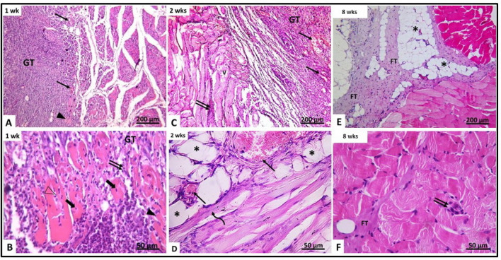
Fig. 4.
H&E-stained section of gluteal muscle from laceration group (group II) at different time intervals. A, B After 1 week (IIa) (A) showing granulation tissue (GT) containing mononuclear inflammatory cells, congested blood vessels (↑) and disrupted muscle fibers (▲). The granulation tissue (GT) contains fragmented muscles with pyknotic nuclei (thick arrow). Other muscles contain neutrophils (Δ). C, B After 2 weeks (IIb) showing disrupted vacuolated muscles (V), congested blood vessels (↑) and inflammatory cellular infiltration (↑↑). D Showing congested blood vessels (↑), fat cells (*) and regenerating myotubes containing centrally located nuclei (curved arrow). E, F After 8 weeks (IIc) showing the area of injury containing fibrous tissue and fat cells (*). Minimal inflammatory cellular infiltration (↑↑) and fibrous tissue (FT) are seen between muscle fibers

