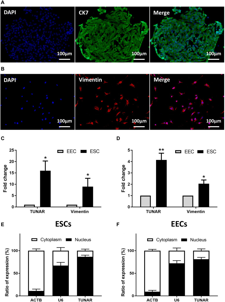
FIGURE 2.
Localization of TUNAR in human endometrium. Immunofluorescence staining of CK 7 (green) in EECs (A) and vimentin (red) in ESCs (B) (200×). The nuclei were stained with 4′,6-diamidino-2-phenylindole (DAPI) (blue). (C) The expression of vimentin mRNA and TUNAR in EECs and ESCs from the same patients in the late proliferative phase (n = 3). (D) The expression of vimentin mRNA and TUNAR in EECs and ESCs from the same patients in LH + 7 phase (n = 3). The subcellular localization of ACTB, U6, and TUNAR in (E) ESCs (n = 3) and (F) EECs (n = 3). *P < 0.05, **P < 0.01.

