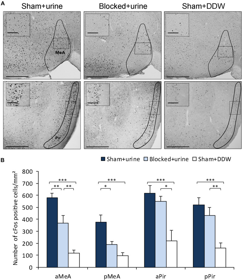
FIGURE 3.

Obstruction of the NPD reduces pheromone-induced neuronal activity in the MeA. (A) Representative images of cFos staining in coronal sections of sham + urine (left panels), blocked + urine (middle panels), and sham + DDW (right panels) mice. Anatomical areas of interest are outlined in black. Insets depict areas outlined by dotted rectangles. (B) Quantification of cFos reactivity in secondary chemosignaling processing brain regions. Blocked male mice exposed to female urine presented decreased neuronal activity when compared to sham mice in the medial amygdala, but not in the piriform cortex. aMeA, anterior medial amygdala; pMeA, posterior medial amygdala; aPir, anterior piriform cortex; pPir, posterior piriform cortex. Scale bar: 500 μm, inset: 100 μm. *p < 0.05, **p < 0.01, ***p < 0.001.
