
Figure 3.
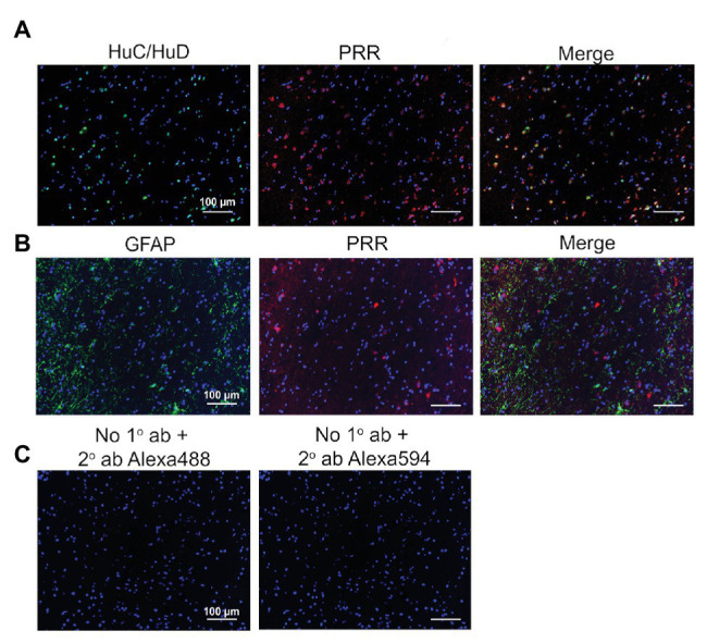
Expression of the PRR in PVN neurons of the human brain. (A) PVN brain tissues were immunolabeled for the PRR (red) and the neuronal marker HuC/HuD (green). (B) PVN brain tissues were immunolabeled for the PRR (red) and the astrocyte marker GFAP (green). (C) Sections processed without primary antibody but with Alexa 488 (green) or Alexa 594 (red) secondary antibody were used as negative controls. ab, antibody; 1°, primary; and 2°, secondary.