
Fig. 3. Donor in vivo differentiated ML NK cells traffic to the BM and are phenotypically similar to PB ML NK cells.
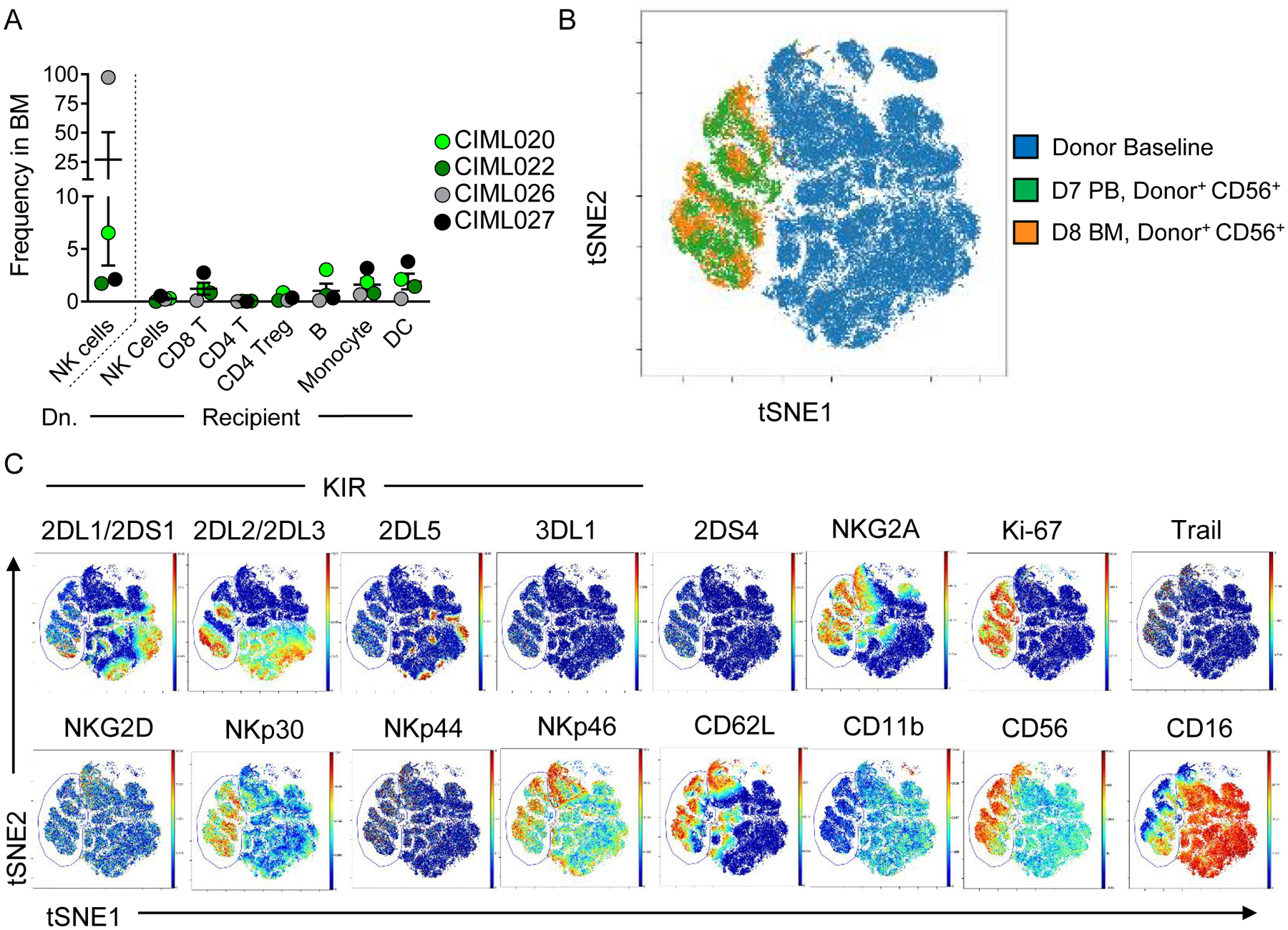
(A) Summary data from Citrus-gated lymphocyte populations in patients with bone marrow (BM) assessed by mass cytometry at day 8 post NK cell infusion. (B) viSNE overlay of donor NK cells at BL (blue), donor NK cells in the PB (green), and donor ML NK cells in the BM, using the same clustering as in (Fig. 1–2). (C) Representative expression of indicated markers in BL NK cells and donor ML NK cells (indicated within the gate) from the PB and BM.