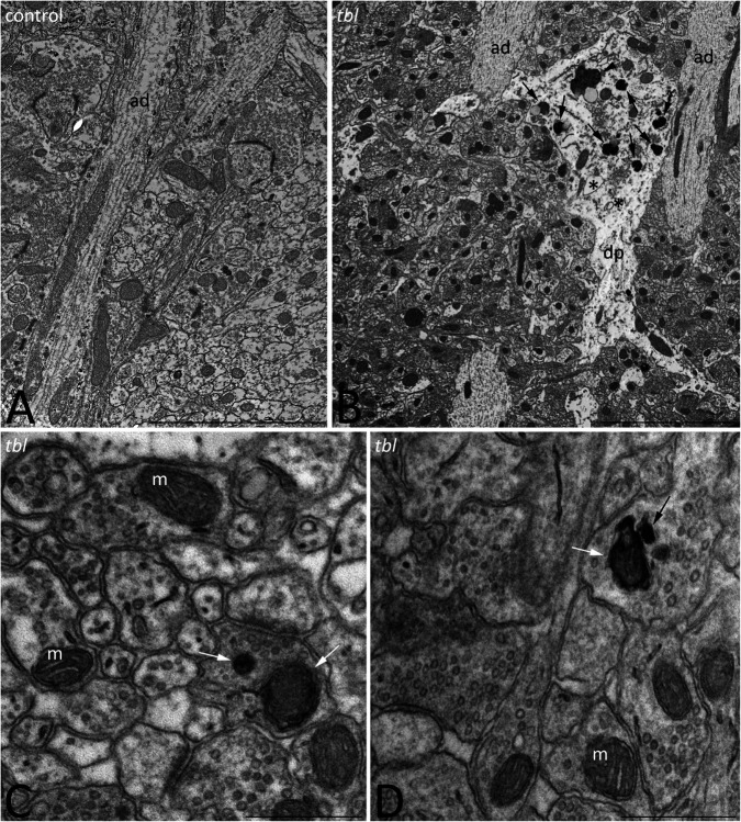
FIGURE 10.
Transmission electron microphotographs through the stratum radiatum in the hippocampal CA1 region of control (A) and tbl (B–D) mice. Degenerative profiles (B, dp) whose cytoplasm is filled with altered vacuoles (B, asterisks), numerous lysosomes and necrotic debris (B, arrows), and mitolysosomes (B, arrowhead) are evident in the tbl mutant CA1. Mitophagic vacuoles and dark debris are often observed in axons and presynaptic terminals throughout the tbl CA1 neuropil (C,D, arrows). ad, apical dendrite; m, mitochondria. Bar = 5 μm (A,B), and 0.5 μm (C,D).

