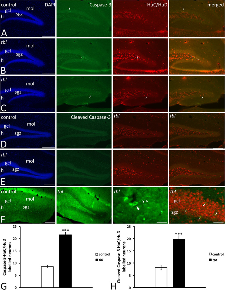
FIGURE 9.

Microphotographs of coronal sections through the rostral (A,B) and middle (C–F) regions of the dentate gyrus of control (A,C,E) and tbl mice (B,D,F) to illustrate the immunoreactivity to caspase 3 and the neuronal marker proteins HuC/HuD. Almost all caspase-3 immunoreactive cells are located within the hilus, the subgranular zone and the granule cell layer, while HuC/HuD also labels interneurons located in the molecular layer. In the control DG it is possible to find caspase-3 labeled cells that do not co-express HuC/HuD (A,C, arrows), whereas in the tbl DG, caspase-3 immunoreactive cells also are labeled by the HuC/HuD antibody (B,D, arrows). Quantitative analysis demonstrates that the tbl DG has significantly more caspase-3-HuC/HuD (G, ***p = 1.902245E–12) and cleaved caspase-3-HuC/HuD double labeled neurons than the controls (H, ***p = 5.524E–06). gcl, granule cells layer; h, hilus; mol, molecular layer; sgz, subgranular zone. Bar = 200 μm (A–F), and 30 and 60 μm (F).
