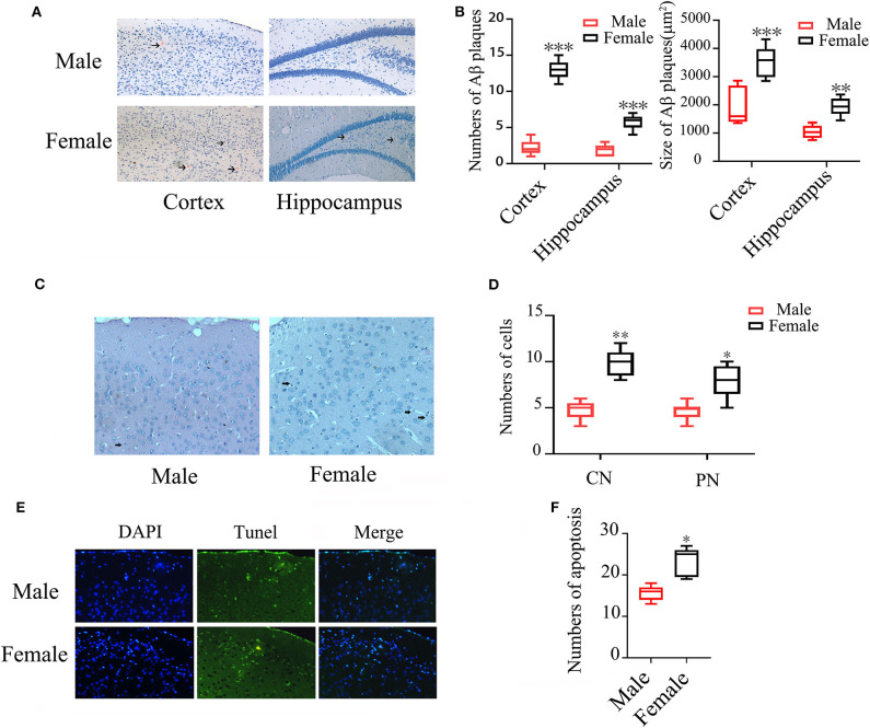
Figure 1.
Aβ plaques and neuronal apoptosis in brains of male and female APP/PS1+/− mice. (A) Congo red staining showing Aβ plaques in the cortex and hippocampus of APP/PS1+/− mice; (B) Quantification of Aβ plaque numbers. (C) HE staining showing condensed (CN) and pycnotic nuclei (PN) (arrows indicated) in cortex of APP/PS1+/− mice; (D) Quantification of the numbers of condensed and pycnotic nuclei; (E) TUNEL staining showing neuronal apoptosis in the cortex of APP/PS1+/− mice; (F) Quantification of the numbers of apoptotic cells. n = 8–10. *P < 0.05, **P < 0.01, and ***P < 0.001 vs. male mice.

