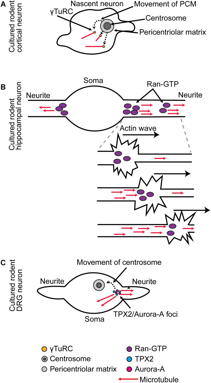
FIGURE 1.
(A) In cultured rodent cortical neurons, as the centrosome is decommissioned as an MTOC, there is a phase of somatic γ-Tub-mediated microtubule organization. (B) In cultured rodent hippocampal neurons, RanGTP localization at the base and distal domains of the extending neurite supports Tpx2-mediated microtubule generation. During neurite outgrowth actin waves that progress along the neurites trigger increased microtubule generation in their wake. (C) In cultured rodent DRG neurons, the centrosome is first situated at the base of one neurite, and at this position Tpx2 facilitates Aurora A activation. As the centrosome migrates away, it leaves behind a new Aurora A-Tpx2 based MTOC.

