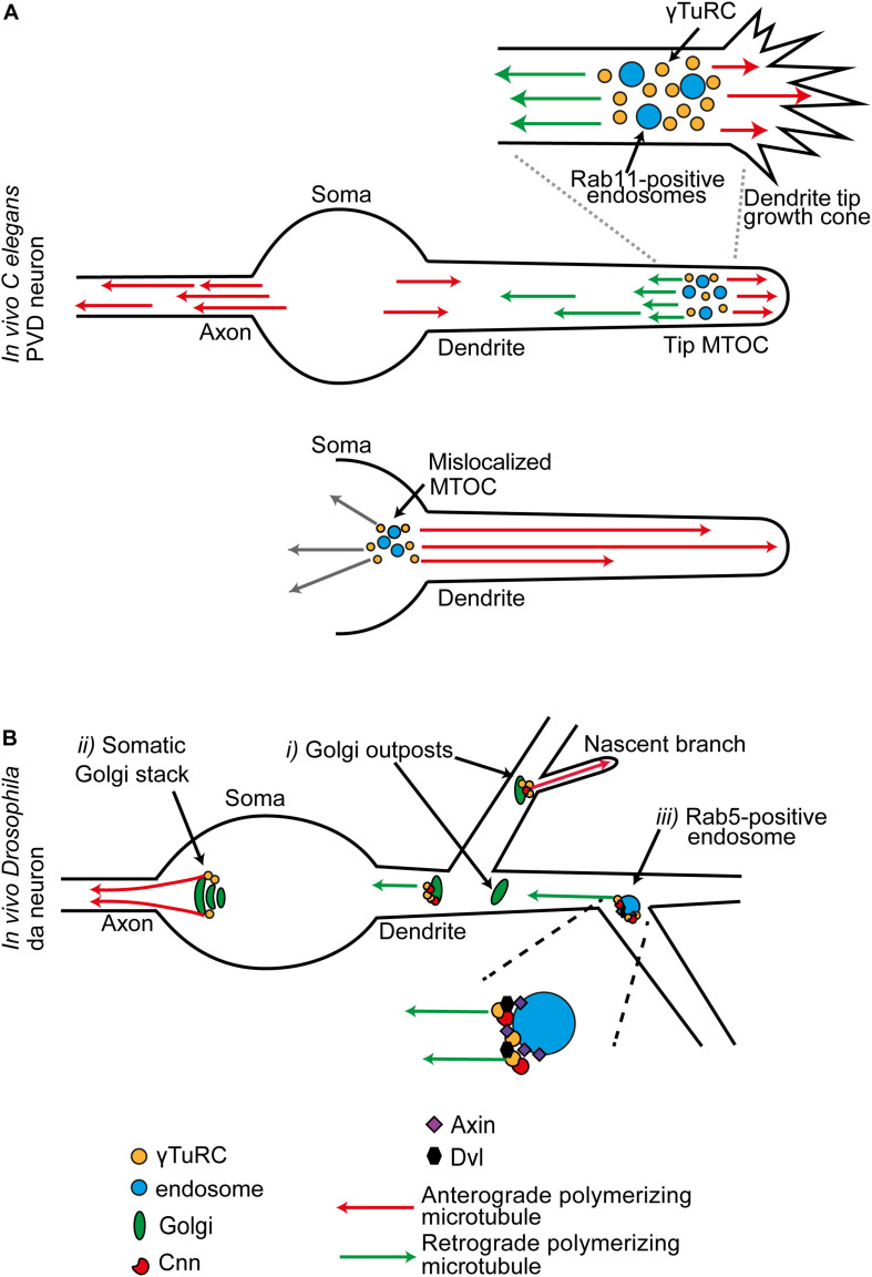
FIGURE 3.
(A) In C. elegans PVD neurons, dendritic growth cone MTOCs give rise to two distinct populations of polymerizing microtubules: plus-ends-out microtubules that invade the tip, and minus-ends-out microtubules that create the specialist polarity organization of the dendrite. This MTOC consists of γ-TuRC organized around a population of RAB11-positive endosomes. When the MTOC is mislocalized to the soma, all microtubules in the dendrite are now plus-ends-out. (B) Sites of MTOC activity in late stage neurons, illustrated for the combined data from multiple studies in Drosophila da neurons. (i) A subset of Golgi outposts is associated with γ-Tub, and unidirectional microtubule generation is promoted from some. (ii) In the mature neuron, Golgi mediated microtubule generation is principally from stacks in the soma. These generate a population of polymerizing microtubules that exclusively invade the axon. Golgi may also act as local site of microtubule generation at the branchpoints of a nascent branch. (iii) γ-Tub is localized at Rab5-positive endosomes at dendrite branchpoints. γ-TuRC-TPs and components of the Wnt signaling pathway are required for microtubule generation activity at these positions.

