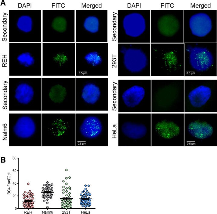
Figure 7.
Evaluation of BG4 foci in various cell lines to determine occurrence of G-quadruplex DNA structures within cells. (A) Evaluation of ability of BG4 binding to G4 structures within different human cell lines, REH, Nalm6, 293 T and HeLa. Cells were fixed in 2% paraformaldehyde and permeabilized with 0.1% Triton-X100, followed by blocking. Immunofluorescence was performed by incubating cells with BG4 at 4°C, followed by anti-FLAG, and anti-rabbit secondary antibody. Following streptavidin-FITC, coverslips were mounted with DABCO/DAPI and images were recorded using Zeiss Apotome, and analyzed with ImageJ. Data were analyzed and plotted as scatter plots using GraphPad Prism. BG4 was tagged with FLAG antibody and observed in FITC channel (green). Nuclei were counterstained with DAPI (blue). First row of every panel is secondary control (only anti-FLAG, anti-rabbit). Second row depicts the representative BG4-stained image. BG4 foci can be observed within cells in the nuclear region, in punctate form. (B) Scatter plots depict the quantification of BG4 foci for REH, Nalm6, HEK293T and HeLa cells.

