Figure 8.
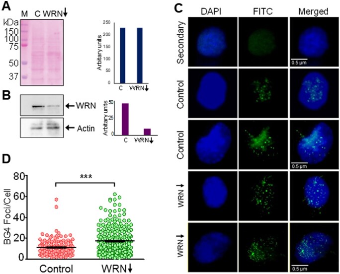
Modulation of G-quadruplex formation within cells following depletion of G4 resolvase, WRN in HeLa cells. (A) shRNA-mediated knockdown of Werner’s helicase, WRN was performed in HeLa. Ponceau-stained blot reveals equal loading of the proteins. (B) Immunoblotting to determine extent of WRN knockdown in cells incubated with shRNA. ACTIN served as loading control and was used for normalization. Bar diagram showing extent of knockdown is also shown. (C) Representative images after immunofluorescence studies showing BG4 foci following WRN knockdown in HeLa cells compared to cells transfected with scrambled shRNA controls. Cells were counterstained with DAPI and scored for G4 structure formation by quantifying BG4 foci, as described above. For other details refer Fig. 7 legend. (D) Scatter plot illustrating the comparison of G4 structure formation determined as a measure of BG4 foci in WRN knockdown cells.

