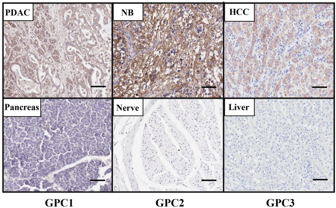
Figure 1.
The protein expression of GPC1, GPC2, and GPC3 in cancers. GPC1 protein level is increased in PDAC tissue compared with normal pancreas as determined by IHC using the GPC1-specific mouse monoclonal antibody (MAb) HM2. GPC2 protein is presented in NB tissue but not in normal peripheral nerve tissue as determined by IHC using the anti-GPC2 mouse MAb CT3. GPC3 is overexpressed in HCC tissue compared with normal liver tissue using the GPC3-specific mouse MAb YP7 by IHC. Images were obtained under 20 × magnification. Scale bar = 200 µm. Abbreviations: HCC, hepatocellular carcinoma; IHC, immunohistochemistry; NB, neuroblastoma; PDAC, pancreatic ductal adenocarcinoma.

