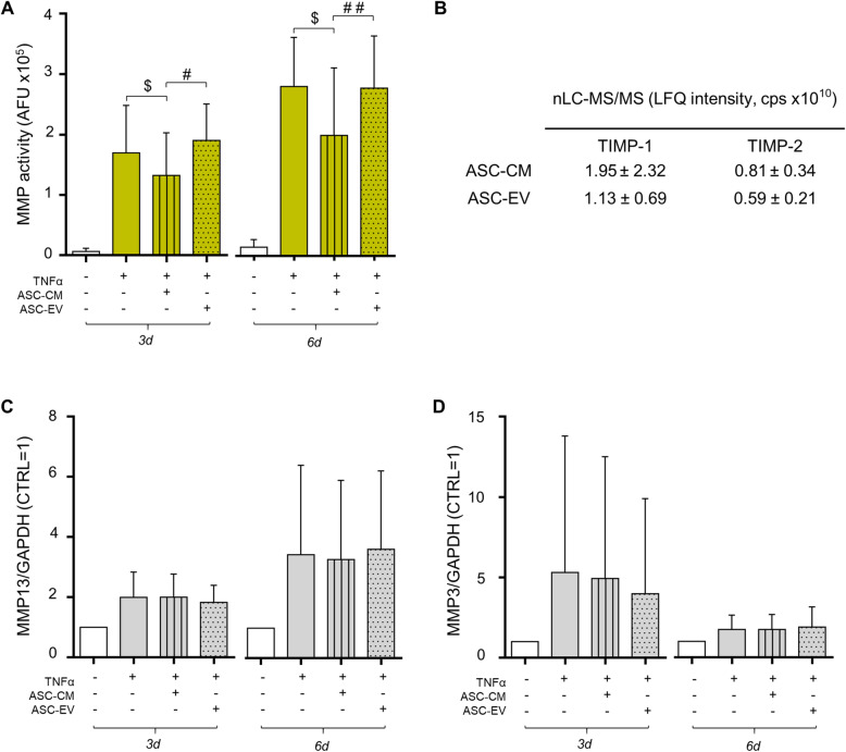
Fig. 2.
Reduction of MMP activity by ASC-CM, TIMP quantification, and MMP expression. a MMP activity, analyzed in CH culture medium (n = 7 independent experiments) 3 and 6 days after the treatments, is expressed as arbitrary fluorescence units (AFU). All conditions statistically differ from control (at day 3: TNF p < .01, TNF+ASC-CM p < .05, and TNF+ASC-EV p < .001; at day 6: TNF p < .001, TNF+ASC-CM p < .05, and TNF+ASC-EV p < .001). Significance vs 10 ng/ml TNFα is shown as $p < .05; vs ASC-EV as #p < .05, ##p < .01. b TIMP-1 and 2 data are expressed as label-free quantification (LFQ) intensity, count per second (cps) from differential proteomic analysis of 20 μg of ASC-CM and -EV proteins. Means ± SD (n = 3) are shown. c, d Quantification of the expression of MMP-13 (c) and MMP-3 (d) in TNFα-stimulated and ASC-CM- or -EV-treated CH at day 3 and 6 analyzed by Western blot. Data (n = 5 independent experiments) were normalized on GAPDH and expressed as relative values (CTRL = 1)

