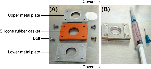
FIGURE 1. Rose chamber for live-cell imaging.
(A) A disassembled Rose chamber to illustrate its components. A Rose chamber contains two metal plates, one 1–3 mm thick silicon rubber gasket, and two coverslips (one that carries the cells and another one that is empty). All components are held together by four bolts. (B) An assembled Rose chamber and a syringe filled with medium. The side of the Rose chamber facing up will be facing the objective during imaging.

