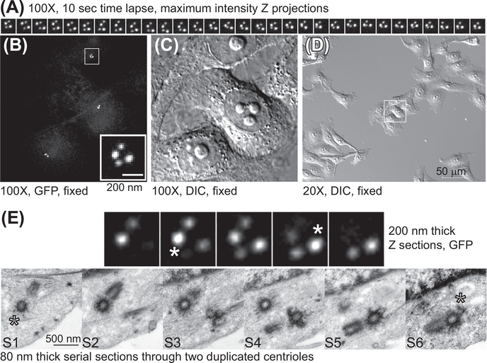
FIGURE 5. CLEM analysis of the centrioles from Centrin1-GFP expressing HeLa cell arrested in G2.
(A) A series of maximal density projections illustrating the movement of the centriole pairs. A Z stack spanning the entire centrosome content was collected using spinning disc confocal and 100× objective lens, every 10 s (B and C) The same cells imaged by time-lapse microscopy were fixed with 2.5% glutaraldehyde and returned to the microscope. The position of the centrioles within the cell was then recorded in fluorescence and DIC. (D) Low-magnification image of the target cell and its neighbor cells in DIC. (E) The centrioles recorded by time-lapse imaged on the electron microscope. Four consecutive immunofluorescence Z sections and six serial EM sections are presented (S1–S6). The asters in S1 and S6 correspond to the immunofluorescence Z section 2 and 4, with maximal intensity for centrin-GFP signal belonging to the mother centrioles (indicating the position of the distal part of the centriole).

