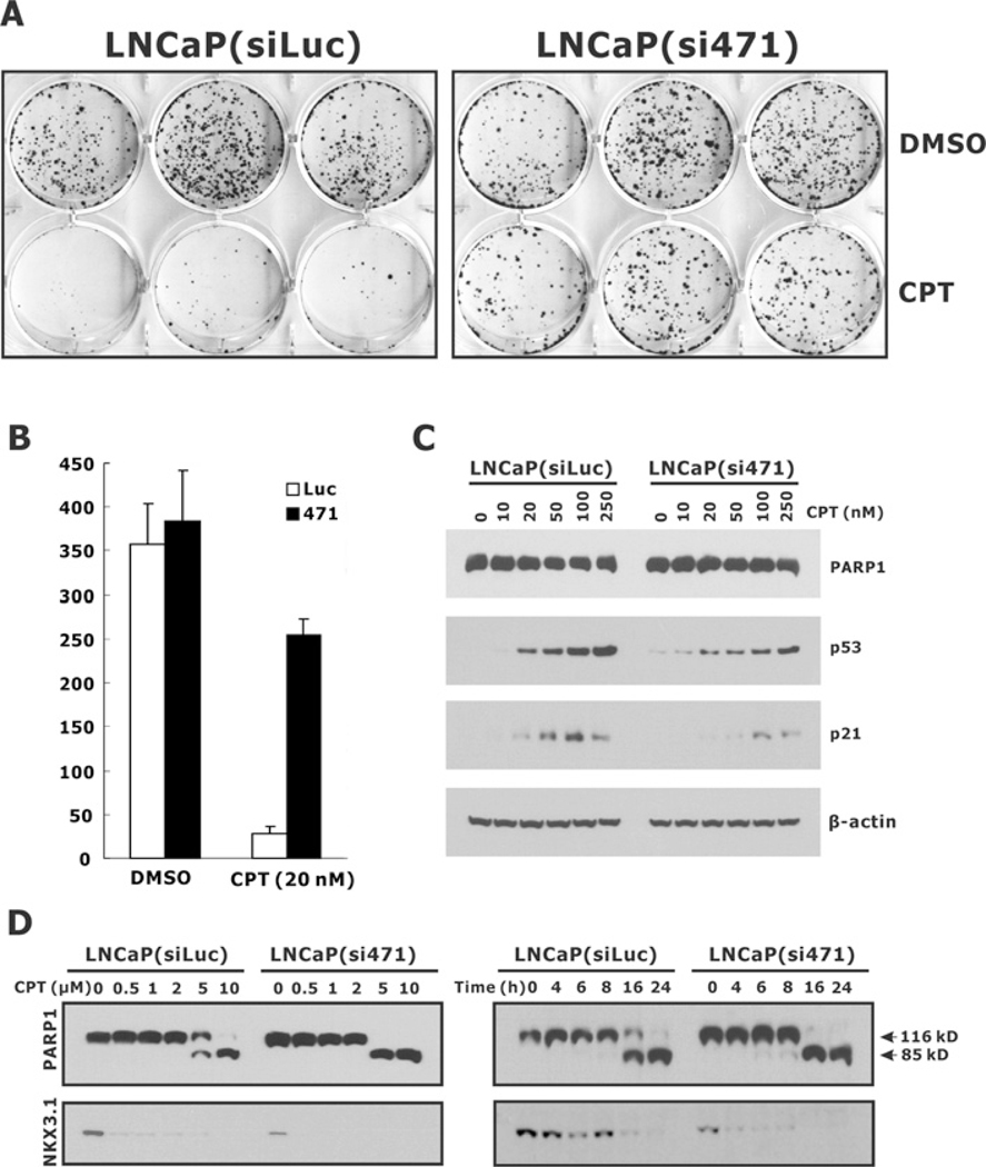
Figure 8. Effect of NKX3.1 knockdown on CPT-induced cytotoxicity in LNCaP cells.
(A) LNCaP(siLuc) and LNcAP(si471) cells were plated into six-well plates at a density of 3000 cells/well and treated with 20 nM CPT. DMSO treatment was used as the vehicle control. Cells were cultured for 2 weeks, stained with Crystal Violet and the colony numbers were counted and plotted (B). (C) LNCaP(siLuc) and LNcAP(si471) cells were treated with increasing amount of CPT for 72 h and the expression of PARP1, p53 and p21 was determined by immunoblotting. (D) Cells were treated with CPT for 24 h (left-hand panel) or treated with 5 μM CPT for the indicated time periods (right-hand panel). The cell lysates were then analysed for PARP1. The intact (116 kD) and cleaved (85 kD) forms of PARP1 are indicated.

