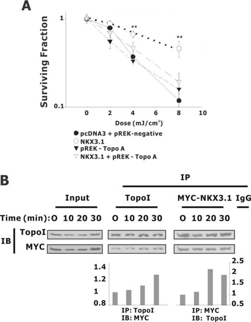
Figure 9. NKX3.1 affects colony formation after the exposure of PC-3 cells to UV irradiation.
(A) PC-3 cells were co-transfected with GFP, pcDNA3 or pcDNA3-NKX3.1, together with the topoisomerase I siRNA vector pREK-Topo A or a control vector pREK-negative. At a density of 2000 cells/well cells were plated into six-well plates and subjected to UV irradiation. Colony formation was counted 7–10 days later and the surviving fraction at each data point was normalized by comparison with colony formation without any treatment. (B) Topoisomerase I (TopoI) association with NKX3.1 in response to UV treatment. HEK-293T cells were transiently transfected with the Myc-NKX3.1 expression vector. Cell lysates were prepared and co-immunoprecipitation (IP) was performed with antibodies against topoisomerase I or Myc. After extensive washing, the immunoprecipitated pellets were fractionated by SDS/PAGE (4–20% gel) and subjected to immunoblotting (IB) with antibodies against topoisomerase I and Myc. The amounts of NKX3.1 (left-hand histogram) or topoisomerase I (right-hand histogram) pulled down were determined by densitometry.

