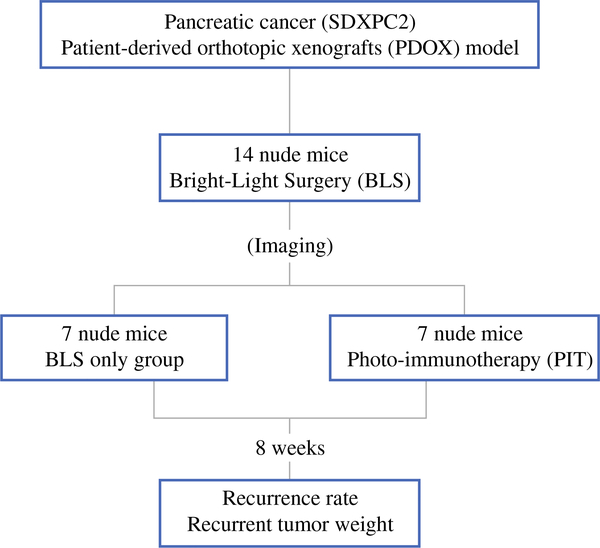
FIG. 1.
Schema of the experimental design. After confirmation of the tumor growth, the PDOXs (SDXPC2) were randomized in the two groups: BLS only and BLS + PIT. Each treatment arm involved seven tumor-bearing mice. BLS was performed under standard bright-field using the MVX10 microscope on both treatment groups. The PDOXs were imaged before and after BLS with the OV100, and the excised tumors also were imaged and weighed. Anti-CEA-IR700 (50 μg) was injected in the tail vain of the mice in the BLS + PIT group 24 h before surgery. An infrared laser (690 ± 5 nm, 150 mW/cm2) was used to irradiate the resection bed of the mice treated with BLS + PIT for 30 min. Eight weeks after surgery, animals underwent laparotomy, and tumors were imaged, weighed, and harvested for analysis

