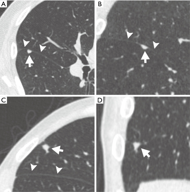
Figure 3.
Samples of typical and atypical perifissural nodules on computed tomography. (A) Axial section showing a typical PFN (arrow) attached to the right major fissure (arrowheads). (B) Coronal section with another typical PFN (arrow) attached to the minor fissure (arrowheads). An atypical PFN (arrow) in axial (C) and coronal (D) sections without visible attachment to the right major fissure (arrowheads in C).

