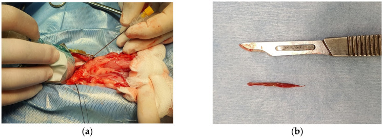
Figure 2.
Photograph of the microconvex probe encased in a sterile protective cover and positioned on the ventral surface of the prostate during the surgery; a spinal needle was introduced, under intraoperative ultrasonographic guidance, through the prostate towards the tip of the awn (a). Photograph of the awn after the removal from the prostate (b).

