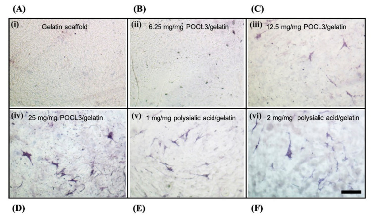
Figure 3.
The alkaline phosphatase staining of murine mesenchymal stem cells (C3H10T1/2) on SBF mineralized gelatin scaffolds. (A) Gelatin scaffold only. (B–D) 6.25, 12.5 and 25 mg/mg POCl3 incorporated with gelatin scaffolds, respectively. (E,F) 1 and 2 mg/mg polysialic acid incorporated with gelatin scaffolds, respectively. The staining showed osteogenic differentiation of stem cells on mineralized scaffolds. Scale bar: 100 μm. Adapted with permission from [11].

