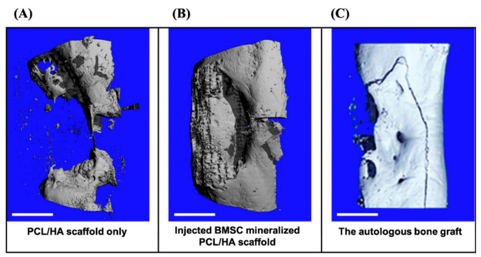
Figure 5.
The micro-CT analysis after 3D reconstructions. (A) The tibia bone defect was repaired with PCL/HA scaffold only. (B) The BMSC cell sheet was injected into the PCL/HA scaffold for in situ mineralization. (C) The autologous bone graft (ABG) positive control. Scale bars: 1 cm. Adapted with permission from [78].

