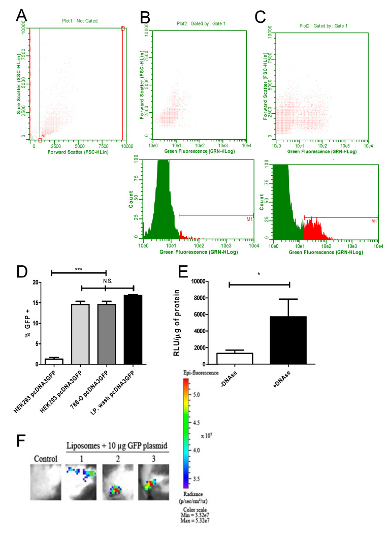
Figure 2.
Efficient transfection of cells with green fluorescent protein (GFP) and luciferase-encoding plasmids encapsulated in cationic liposomes. To analyze the transfecting capacity of DNA-loaded cationic liposomes, HEK293T and 786-O cells were transfected in vitro as described in the Methods section. BALB/C mice were inoculated i.p. with encapsulated pcDNA3-GFP. Then, 24 h after transfection, GFP fluorescence was measured in trypsinized cells or peritoneal cells via cytometry. The gates used for size and side scattering analyses are shown in (A). In (B), cells exposed to empty liposomes (no GFP) are shown, in (C), cells transfected with encapsulated pcDNA3-GFP are depicted. The upper panel shows the dot plot while the lower panel depicts the histogram from the same sample analysis (HEK293 cells used in this experiment). In (D), the results of three independent experiments for each group are summarized, comparing the in vitro transfection from HEK293T and 786-O with the in vivo transfection (cells washed from the peritoneal region 24 h after transfection). HEK293T cells transfected either with encapsulated pcDNA3-GFP (second bar from the left), encapsulated pcDNA3-GFP transfected 786-O cells (third bar from the left) or washed, in vivo transfected peritoneal cells (bar on the right) showed similar fluorescence, significantly different from the HEK293T control (transfected with naked pcDNA3-GFP, left bar, significance was tested with ANOVA test, *** = p < 0.005). In (E), the influence of pcDNA3-luc DNA on the outside of cationic liposomes is shown after transfection, submitting cationic liposomes after loading to DNAse 1 treatment or not. Note that liposomes treated with DNAse showed a stronger transfection capacity, which is seen as a higher light emission mediated by luciferase expression. Relative light units were normalized as Relative Light Unit/µg. This experiment was done in three different samples in triplicate, analyzed by a paired T test with * = p < 0.05. (F) To validate the intradermal route for immunization, animals were tattooed with cationic liposomes loaded with pcDNA3-luc and control animals were tattooed with pcDNA3-PfRH5. The transfection efficiency was analyzed in an IVIS equipment 24 h after the tattooing process. The epi-fluorescence quantification of all (n = 3 per group) animals was measured as 5.24 × 10⁸ with a standard deviation of 1.5 × 10⁸.

