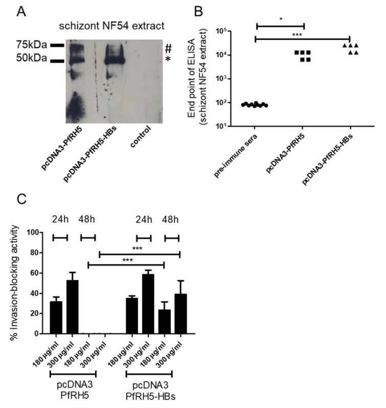
Figure 3.
Antibody response after intradermal tattooing with plasmids encoding PfRH5 (P. falciparum reticulocyte binding protein homologue 5) or PfRH5 fused to small hepatitis B virus envelope antigen (HBs). (A) Sera from pcDNA3-PfRH5 and pcDNA3-PfRH5-HBs immunized mice were used to recognize native PfRH5 in extracts from P. falciparum NF54 schizonts. As a control, pre-immune sera from animals were used. Note that both the processed (*) and unprocessed (#) PfRH5 species were detected. Serum pools diluted 1:500 were employed in all experiments. In (B), endpoint ELISAs were done to measure antiPfRH5 IgG titers in pcDNA3-PfRH5 and pcDNA3-PfRH5-HBs-immunized mice. While pcDNA3-PfRH5-HBs showed values with a mean of 20480 s.d. +/− 7011, animals immunized with pcDNA3-PfRH5 showed values of 10240 s.d. +/− 3505. Assuming equal variance and non-parametric values, a statistically significant difference was observed between immunized-group sera and pre-immune sera (Kruskal–Wallis Test, * = 0.05, *** = 0.0004). In (C), the inhibitory activity of IgGs from antisera was monitored in in vitro growth assays. Data are shown as the percentage of growth inhibition of parasite proliferation (measured by flow cytometry) by purified IgG from immunized mice. A dose dependent assay with controls for each column was used. As controls, cultures grown in the presence of pre-immune sera pooled from each group at the maximum quantity were used (300 µg/mL). The inhibition was calculated as the decrease of parasitemia compared to the control sera-treated cultures, as described in the Methods section. The inhibitory effect of pcDNA3-PfRH5-HBs immunized mice sera after 48 h were significantly higher than that of pcDNA3-PfRH5 mice sera (two-way ANOVA with Bonferroni post-tests), while no statistically significant differences were found between all other groups. The values for 24 h and 48 h treatment are shown. As before, IgG from five animals per group were used.

