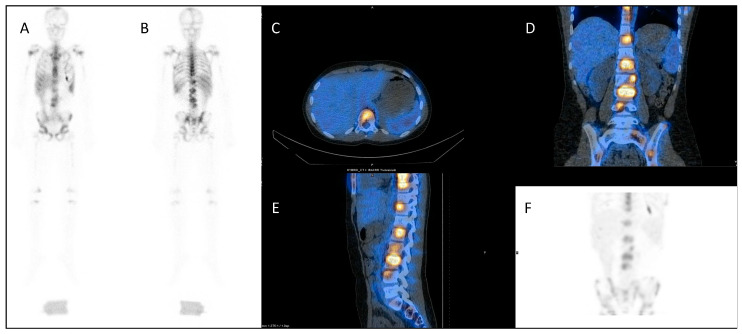
Figure 4.
Dosimetry imaging with 111In-labelled anti-CD66 monoclonal antibody in a patient with relapsed leukaemia prior to 90Y anti-CD66 monoclonal therapy in an early phase trial [50]. (A) Anterior and (B) posterior whole-body planar scintigraphy views; (C) axial, (D) coronal and (E) sagittal fused SPECT CT views; and (F) SPECT maximum intensity projection image. These demonstrate avid uptake in areas of involved bone marrow.

