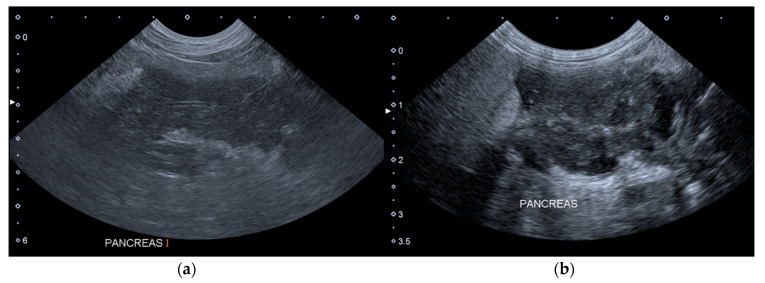
Figure 1.
Longitudinal ultrasound images of the right lobe of the pancreas of a dog included in the group A. (a) T0: the pancreas is enlarged, hypoechoic, slightly inhomogeneous and with peripancreatic hyperechoic mesenteric fat. (b) T1: the pancreas is more inhomogeneous and with peripancreatic free fluid. T0: abdominal ultrasound examination performed on the first day of hospitalization; T1: abdominal ultrasound examination performed on the third day of hospitalization.

