Figure 5.
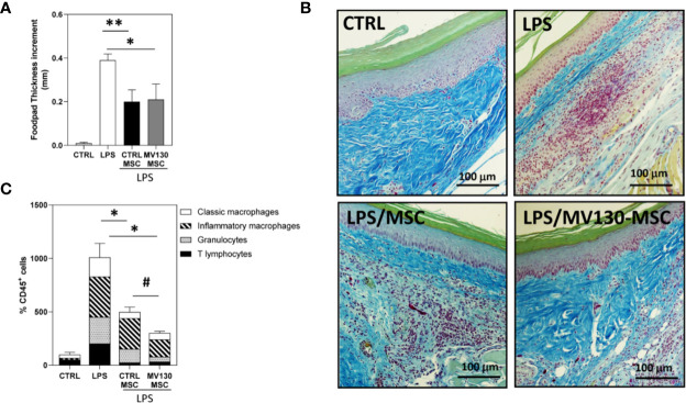
Effects of MV130-MSCs in an in vivo model of acute inflammation. FVB/NJ mice were challenged in the footpad with 40μg of LPS and administered with or without control or MV130-primed MSCs 24 h later. (A) Footpad thickness increment was determined after 72 h as a measure of the efficacy of the different experimental groups of MSCs. Data shown are mean ± SEM of two independent experiments (three mice per group) (B) Images show histological sections of footpad tissue stained with Gallego’s Trichrome. Images are representative of 3 mice per group. (C) Percentage of CD45+ leukocytes infiltrating footpads in the different mouse groups analyzed by flow cytometry. The distribution of the different leukocyte subpopulations in CD45+ cells is also shown in each experimental group. Results represent increments relative to control animals (2 independent experiments with 3 mice per group) (*p < 0.05,**p < 0.01 versus LPS alone; #p < 0.05 versus CTRL-MSCs; by Wilcoxon test).

