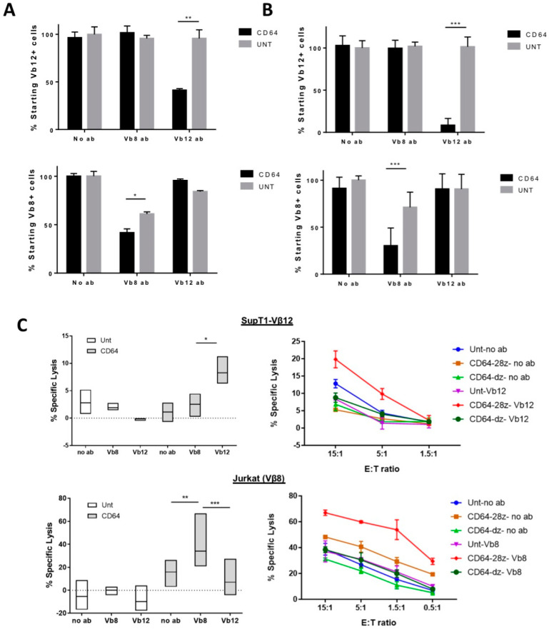
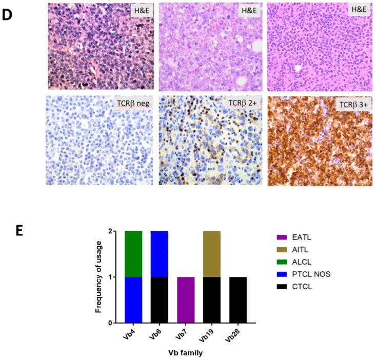
Figure 2.
CD64 IR-modified T cells display specific cytolytic function against TCR Vβ families. (A,B) Autologous lysis of peripheral blood Vβ12 and Vβ8 T cells at 24 h. Vβ-family-specific antibodies were used to “pre-arm” effector T cells (A) and “pre-paint” target T cells (B). After being co-cultured for 24 h, cells were stained with TCR Vβ-directed antibodies analyzed by flow cytometry to determine the relative change in target cells compared to the starting number. TCR Vβ-specific lysis was observed when CD64-IR cells were redirected against both Vβ8 and Vβ12 cells with both methods. (C) Co-culture of T cells and SupT1-Vβ12 and Jurkat T cell line (Vβ8 family) and chromium release assay was performed at 4 h. Left: E:T ratio was 5:1. Data shown for 4 experiments and are normalized to antibody-only control. Right: 4-h chromium release assay performed at different E:T ratios. Technical replicates shown. TCR Vβ-specific lysis was observed when CD64-IR cells were redirected against both Vβ8 and Vβ12 cells. (D) Representative H&E staining (top) and T cell antigen receptor βF1 antibody staining (bottom) of paraffin embedded sections of T cell lymphomas. βF1 antibody staining shows cell surface expression of α/β TCR. Intensity of staining ranged from negative to 3+. (E) Vβ family expression in eight assessable T cell lymphoma patient specimens shows highly varied TCR Vβ family usage. Vβ family usage is determined by TCRβ NGS, and protein expression is determined by immunohistochemistry (βF1 antibody). Asterisk coding in figures is as follows: * p ≤ 0.05; ** p ≤ 0.01; *** p ≤ 0.001.


