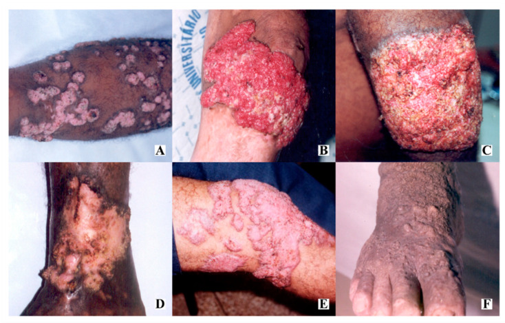
Figure 1.
Pattern of dermatological lesions of chromoblastomycosis: (A) nodular lesions, sometimes confluent; (B) tumoral and hypervascularized lesion with spontaneous bleeding; (C) warty plaque; (D) scar lesion with central healing areas; (E) polymorphic lesion, with nodules, plaques and tumor; (F) verruciform lesion.

