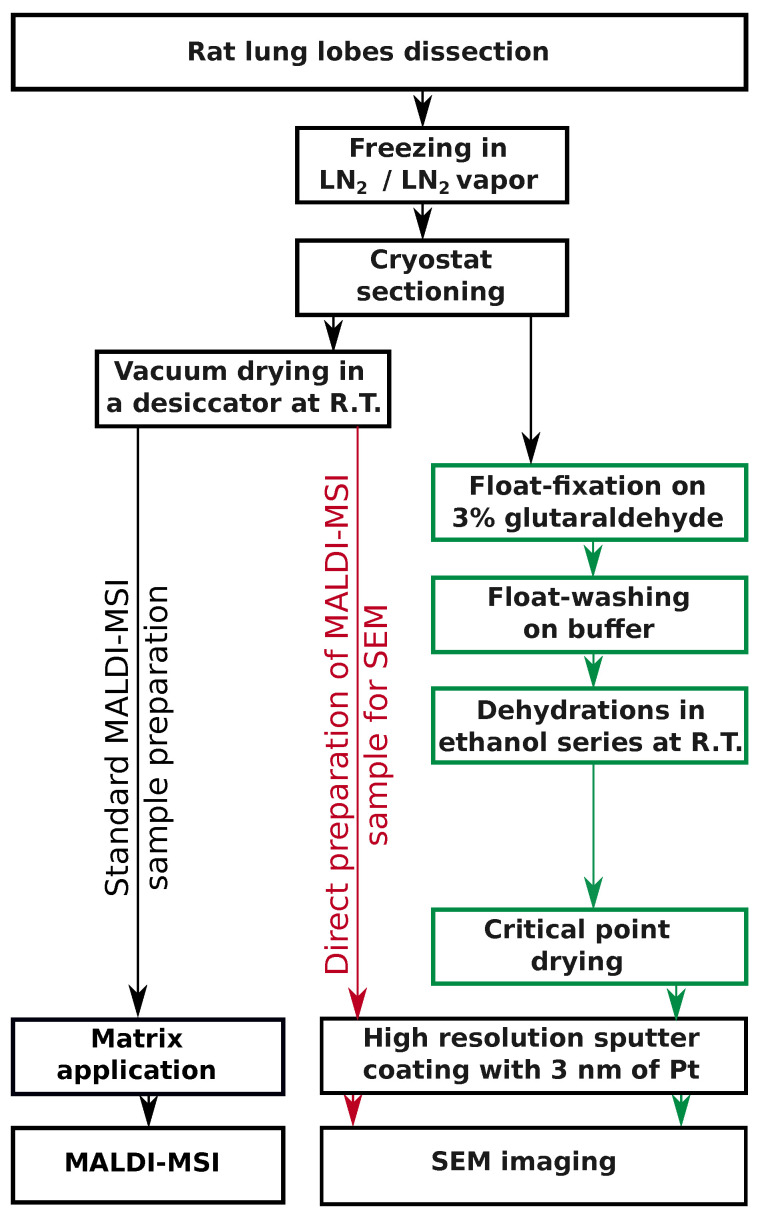
Figure 1.
An overview of lung sample processing for SEM and MSI. The individual steps of the float-fixation/washing procedure are shown in green. MALDI-MSI samples directly prepared for SEM are enhanced in red. Additionally, parallel processing of frozen vacuum-dried lung tissue sections for MALDI-MSI analysis is shown in black in the left part of the panel. Color coding was selected according to AAAS color palette [34]. LN, liquid nitrogen; R.T., room temperature.

