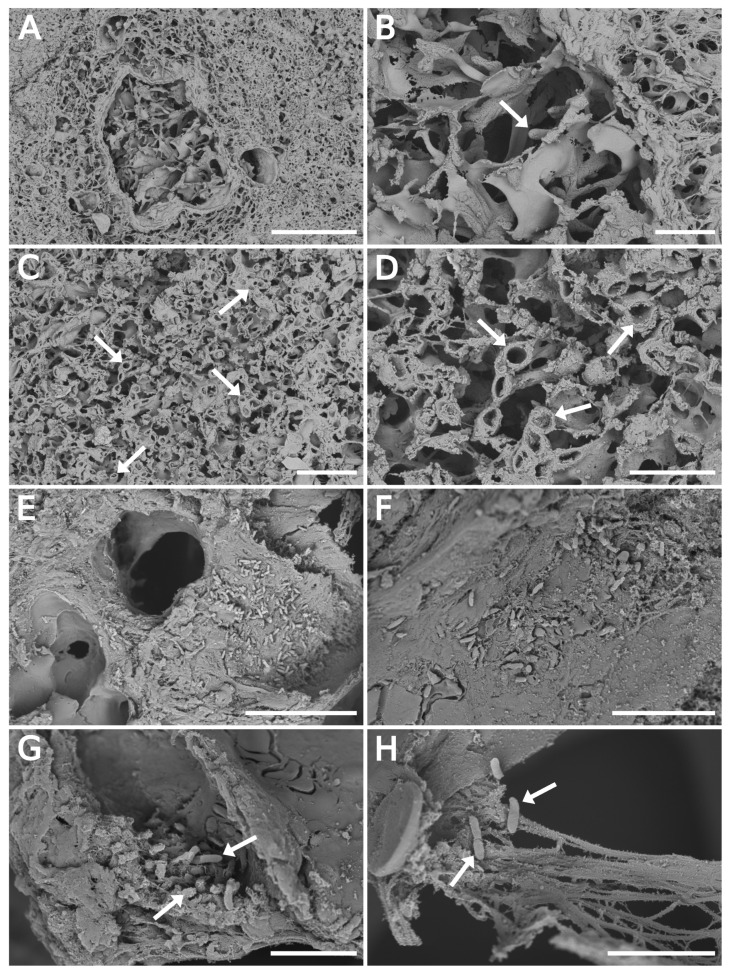
Figure 3.
SEM backscattered electron imaging of float-fixed cryosections from rat lung lobes. (A–D) Lung tissue infected with A. fumigatus, 100 -thick cryosections. (A,B) Fungal hyphae invading the pulmonary parenchyma. The arrow points to the hyphae tip. (C,D) Cross-sectioned hyphae (arrows) in the alveolar space. (E–H) Lung tissue infected with P. aeruginosa, 50 -thick cryosections. (E,F) Overview images of alveolar space with bacteria. (G,H) Bacteria in damaged tissue. Arrows point to individual bacterial cells. Primary magnification of SEM images were: (A) 1000×; (B,C) 3500×; (D) 10,000×; (E) 6500×; (F) 12,000×; (G) 20,000×; and (H) 25,000×. Scale bars: 100 (A); 20 (B,C,E); 10 (D,F); and 5 (G,H).

