Figure 3.
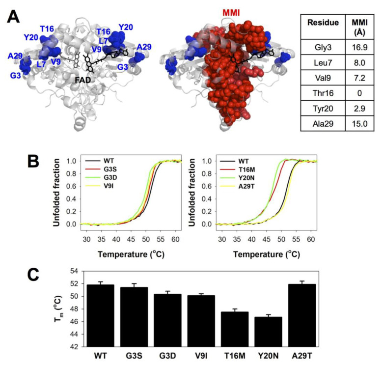
Thermal stability of NQO1 variants. (A) Structural location of the mutated residues, showing their proximity (as a minimal distance) to the monomer:monomer interface (MMI). The structure used for display has the PDB code 2F1O [72]. Residues belonging to the MMI were identified as described [90]. (B) Thermal denaturation profiles; (C) Tm values (mean ± s.d. from six replicas, using proteins from two purifications).

