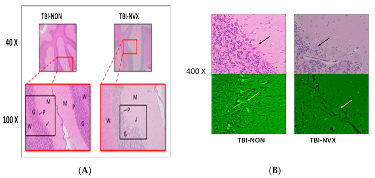
Figure 7.
(A) Histopathology of cerebellar brain tissue. H&E stain from 40× to 400×. Red rectangles delineate the 40× areas that are further magnified by 100×. Black rectangles delineate the areas which are magnified by 200×. G = granular cell layer, M = molecular cell layer, p = Purkinje cell layer, and W = white matter; shrunken, pyknotic, and eosinophilic staining of the Purkinje cells from the TBI-NON animal. Black arrows point to the Purkinje cells further magnified at 400× in (B); (B) High magnification (400×) of framed panel in (A); cerebellar brain tissue using H&E (top) and Fluoro-Jade B (FJB, bottom). The FJB stains at 400× magnification are from adjacent sections to the respective H&E stains above them. Yellow arrow (FJB) points to adjacent Purkinje cells; note the fluorescent staining of the Purkinje cells in the TBI-NON animal and not in the TBI-NVX.

