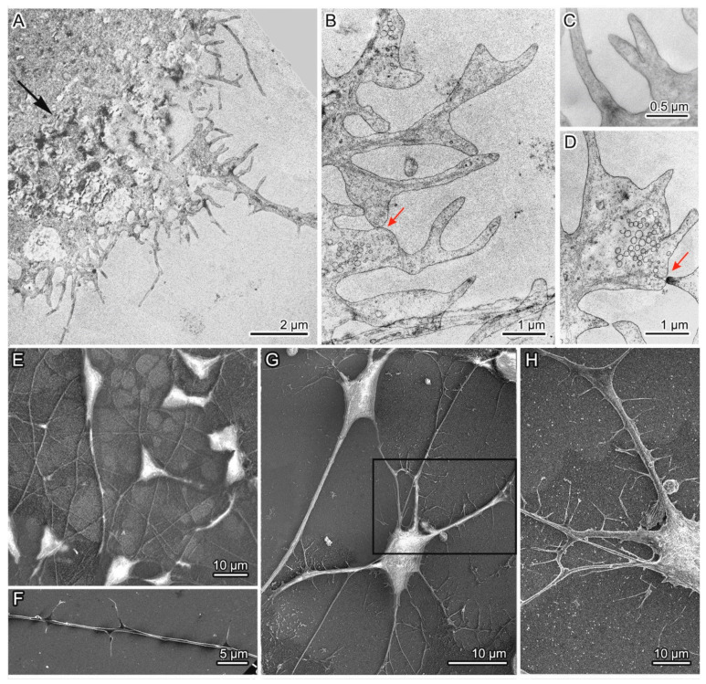
Figure 9.
Abnormal organization of some neurons among group 2 mutant neurons: (A) A part of an atypical-morphology neuron containing a dense aggregate in the cytoplasm (indicated by a black arrow) and a large number of short and long dendrites with spines randomly located on them; (B,C) Spines with a defective shape: bifurcation or adhesion of closely spaced structures; (D) A contact of defective spines and a synapse of abnormal organization; (E,F) Neurons and a dendrite with spines in the control culture; (G) A mutant neuron from group 1 (left) and an atypical neuron from group 2 (right) with dendrites and spines; (H) An enlarged part of an atypical neuron showing a large number of spines on the dendrite. The same fragment is highlighted in the frame in the panel G. Images in panels A–D were obtained by TEM, and in panels E–H via SEM. Contacts of spines and synaptic terminals are indicated by red arrows.

