Figure 2.
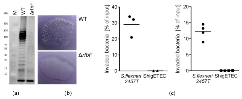
(a) SDS-PAGE gel image of separated LPS from Shigella flexneri 2457T wild-type (WT) and Shigella flexneri 2457TΔrfbF mutant following staining with Pro-Q® Emerald 300 Lipopolysaccharide Gel Stain Kit. The lowest band represents the lipid A-core molecules, while the upper ladder-like pattern is the LPS molecule with various number of O-antigen repeating units. (b) Agglutination assay with Shigella flexneri 2457T wild-type (WT, top panel) and its isogenic ΔrfbF mutant (bottom panel) using rabbit anti-Shigella flexneri 1–6 serum. (c) HeLa cells were infected with the wild-type parental Shigella flexneri 2a 2457T or ShigETEC at a MOI of 80. Percentage of intracellular (invaded) bacteria relative to the inoculum was determined by CFU calculations after plating. Data are shown from two independent experiments.

