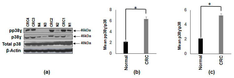
Figure 1.
Increased protein expression and activation of p38γ in human cancerous colonic tissue compared to normal subjects. (a) Whole tissue extracts from colon cancer patients and normal subjects were prepared and 25 µg of protein subjected to SDS-gel electrophoresis and Western blot analysis using antibodies against p38γ and pp38γ. Equal loading was confirmed using an antibody against total p38 antibody. Representative blotting is shown for four normal (N) and four colon cancer (CRC) subjects. (b) Densitometric values expressed as fold increase of ratio of phosphorylated p38γ/total p38. (c) Densitometric values expressed as fold increase of the ratio of p38γ/total p38. The data was analyzed using the paired, two-tailed Student’s t-test, and the results were expressed as fold change ± SEM of 10 colon cancer patients and 10 normal subjects * p-value < 0.001.

