Figure 4.
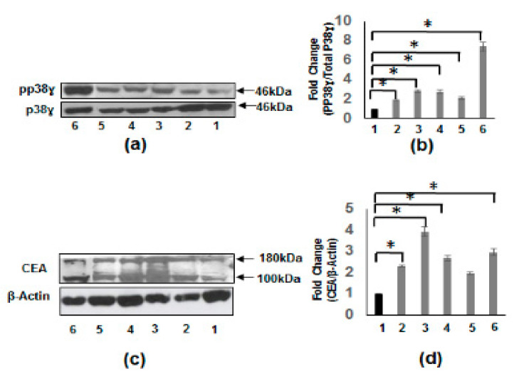
Increased phosphorylation of p38γ and protein expression of CEA in CRC cell lines. (a) Western blot using specific antibodies to p38γ and pp38γ in cell extracts from IEC-6 rat intestinal epithelial cells (lane 1), HCT 116 human CRC cells (lane 2), HT-29 human CRC cells (lane 3), Caco-2 human CRC cells (lane 4), SW620 human CRC cell line (lane 5), and HepG2 human hepatoblastoma carcinoma cell line (lane 6). (b) Densitometric values expressed as fold increase of the ratio of pp38γ/p38γ mean ± SEM (n = 4) * p-value < 0.05. The above results show the increased phosphorylation in CRC cell lines and HepG2 cells compared to normal IEC-6 cells. (c) Whole cell extracts from different cell lines were subjected to Western blotting with specific antibodies to CEA. (d) Densitometric values showing the fold increase of the ratio of CEA/β-actin mean ± SEM (n = 4) * p-value < 0.05. As shown in (c) the CEA levels are uniformly increased in colon cancer cell lines and HepG2 cell line.

