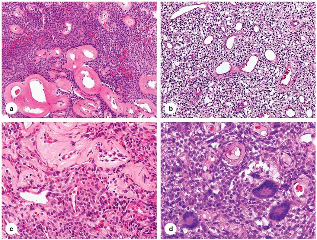
Figure 1.

Histological features of glomangiopericytoma. (a–c) The tumor is composed of epithelioid cells set in a perivascular pattern around prominent blood vessels with prominent perivascular hyalinization. (d) One example contained multinucleated giant cells.
