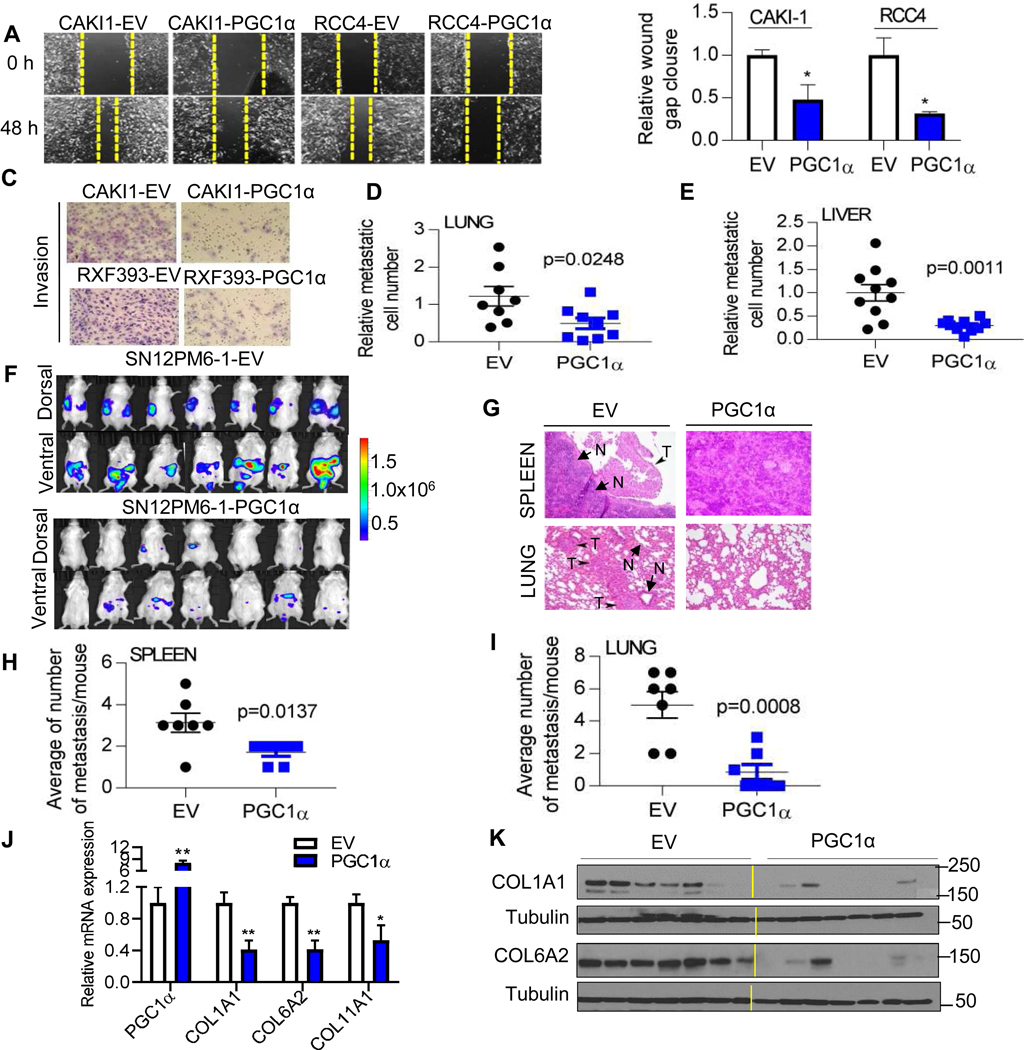
Fig. 3. PGC1α suppresses the migratory and invasive phenotypes of ccRCC cells.
(A) Representative images and (B) relative wound healing over time in RCC cells (n=3/group). (C) Representative images of Boyden invasion assay with Matrigel insert in RCC cells. (n=3/group). (D and E) Chick embryo chorioallantoic membrane (CAM) metastasis assay. Two million RXF393 cells were implanted on the upper CAM vessels of 10-day old chick embryos. One week later, tissues were harvested and metastasis of RCC cells was quantified by qPCR of human Alu repeats in genomic DNA (n=10/group). (F) Bioluminescence in vivo imaging at 6 weeks after tumor challenge. SN12PM6-1 cells were orthotopically implanted into the left kidney of SCID mice (n=7/group). (G) H&E staining of tissue sections from the mice orthotopically implanted with an EV or PGC1α expressing SN12PM6-1 cells at 6-week after tumor challenge. Arrows indicate metastatic tumors (T) and normal tissues (N). Spontaneous metastasis to the spleen (H) and lung (I) was quantified. (J) At 6 weeks from tumor challenge, kidney tumors were harvested and analyzed for the mRNA expression of PGC1α and COLs (n=7/group). (K) COL1A1 and COL6A2 protein levels in kidney tumors from the mice orthotopically implanted with an EV or PGC1α expressing SN12PM6-1 cells (n=7/group).

