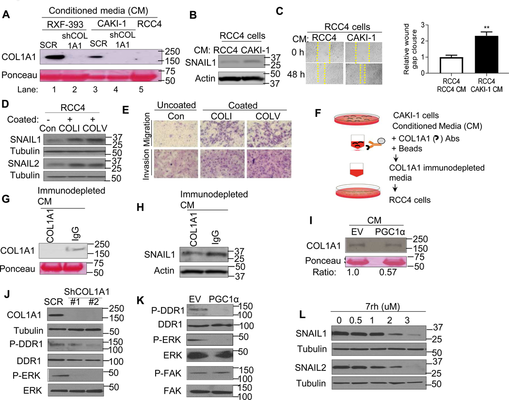
Fig. 6. PGC1α suppresses SNAIL protein via deactivation of collagen-induced DDR1 axis.
(A) Western blot analysis of COL1A1 in the conditioned media (CM) from RCC cells stably expressing shRNA control (SCR), COL1A1 shRNA, or RCC4 cells. (B) Immunoblot analysis of SNAIL1 expression in RCC4 cells cultured with the CM from RCC4 or CAKI-1 cells. (C) Representative images and quantification of migratory phenotype in RCC4 cells cultured with the CM from RCC4 or CAKI-1 cells (n=3/group). (D) Western blot analysis of indicated proteins in RCC4 cells cultured on a plate pre-coated with either collagen type I (COLI) or collagen type V (COLV) for 72 h. (E) Representative images of migratory and invasive phenotype in RCC4 cells cultured on a plate pre-coated with either collagen type I (COLI) or collagen type V (COLV) for 72 h (n=3/group, 3 independent experiments). (F) Illustration of COL1A1 immunodepletion assay. The conditioned media (CM) from CAKI-1 cells were incubated with either COL1A1 antibodies or IgG coated magnetic beads. The COL1A1 immunodepleted media were incubated with RCC4 cells. (G) Western blot analysis of COL1A1 in the CM from COL1A1 immunodepletion or IgG control pull down. (H) Western blot analysis of SNAIL1 expression in RCC4 cells cultured with the CM from COL1A1 immunodepleted media or IgG control pull down. (I) Western blot analysis of COL1A1 in the CM from CAKI-1 cells stably expressing an EV or PGC1α. (J) Western blot analysis of indicated proteins in CAKI-1 cells stably expressing shRNA control (SCR) or two independent shRNA COL1A1. (K) Western blot analysis of indicated proteins in CAKI-1 cells stably expressing an EV or PGC1α. (L) Immunoblot analysis of indicated proteins in CAKI-1 cells treated with pharmacological DDR1 inhibitor 7rh for 24 h.

