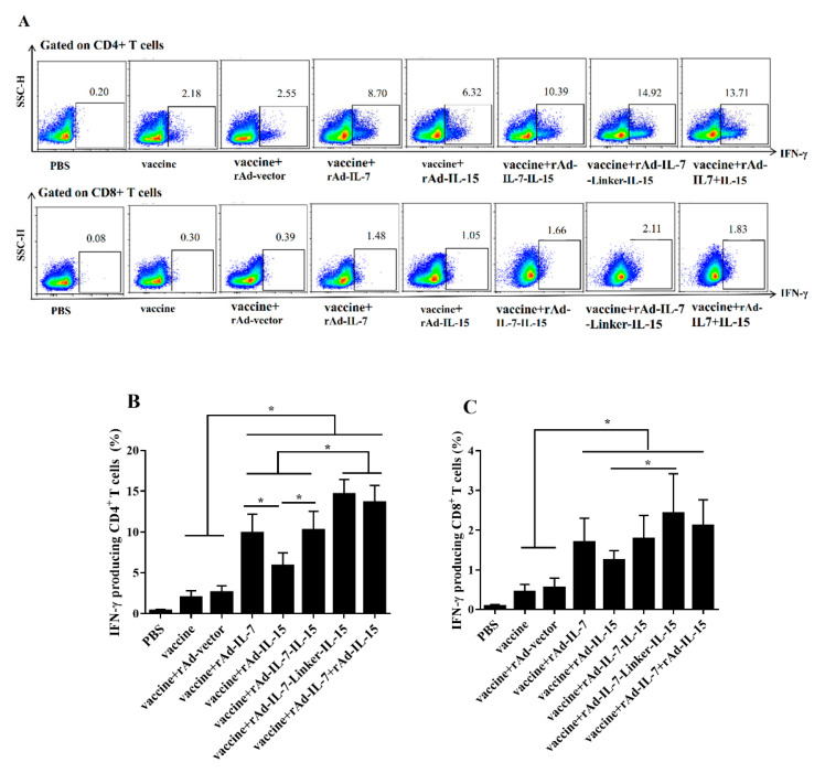
Figure 2.
IFN-γ secretion following twice stimulation with antigens in spleens. At 20 weeks after the last immunization, mice were injected with BCG (1 × 10⁶ CFU) by i.p for 9 days. Then, lymphocytes of spleens were isolated and stimulated with a cocktail of antigens ESAT-6, Ag85B, Rv2626c and HspX (2 μg/mL) in vitro. Secretion of IFN-γ was determined by flow cytometry. (A) The representative results of every group; (B) CD4+ T cells secreting IFN-γ; (C) CD8+ T cells secreting IFN-γ. Data collected were presented as means ± SD from 5 mice per group. * p < 0.05.

