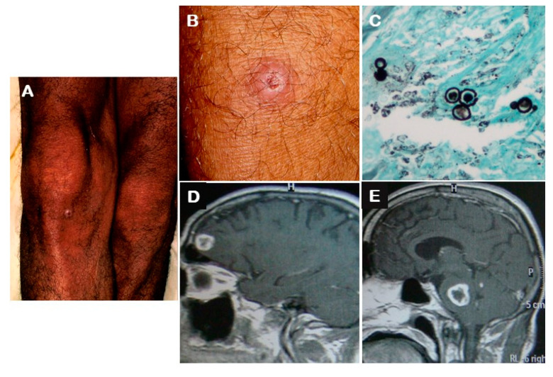
Figure 1.
Neuroparacoccidioidomycosis in a patient with the chronic form of the mycosis: (A) A small round erythematous nodular skin lesion (around 1 cm) with central depression covered by crust on the right knee of the patient described as case 1; (B) Closer view of the lesion described in panel A; (C) Histopathological examination (Grocott silver stain) of a fragment of the skin lesion on the knee showing large budding yeast cells of Paracoccidioides sp.; (D) Magnetic resonance image showing a hyperintense (T1) round lesion with peripheral contrast enhancement and surrounding edema on the left frontal lobe (2 cm); (E) A similar brain lesion on the pons (2.5 cm).

