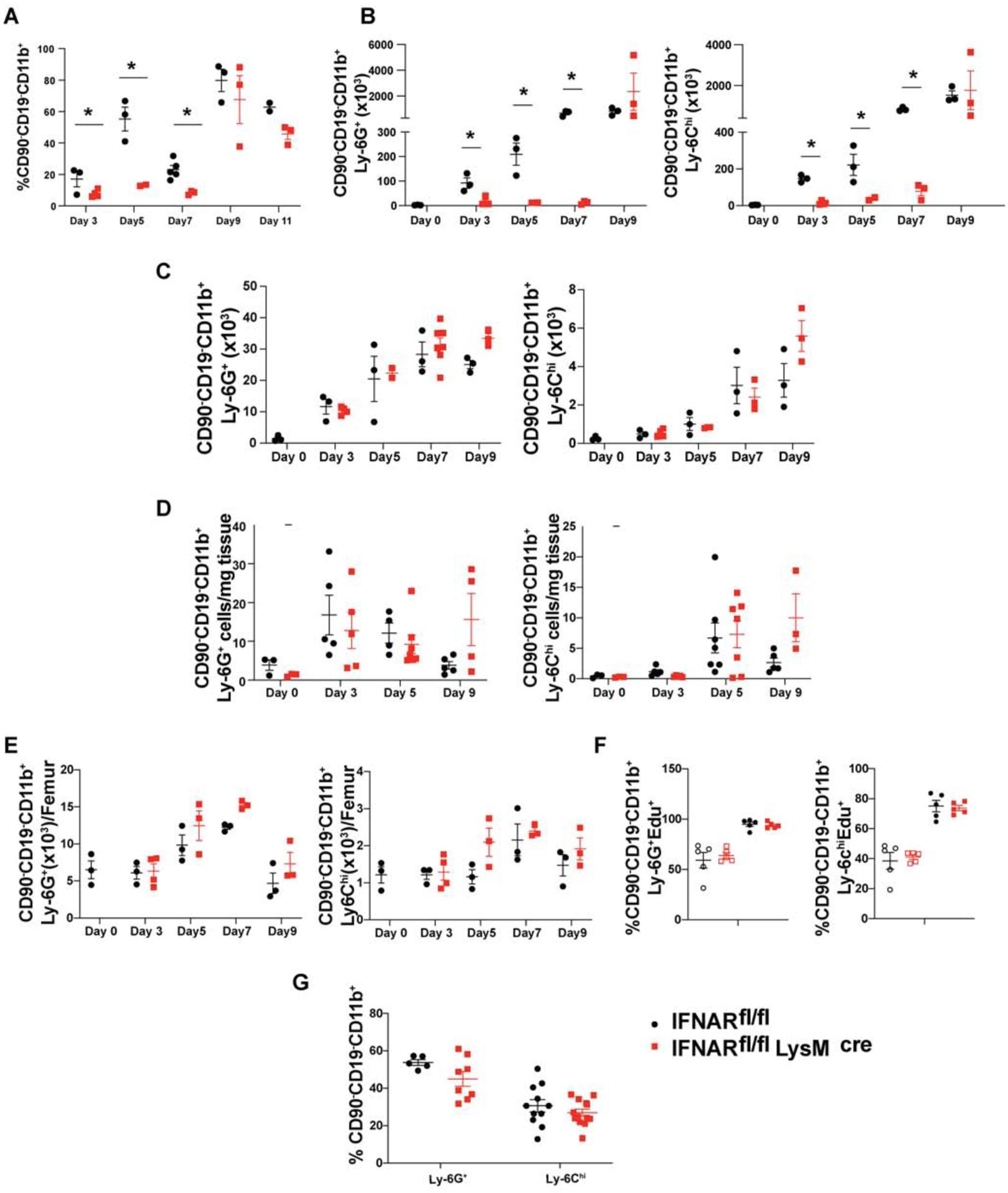
Fig. 4.

Lack of IFNAR signaling on Tregs leads to defective recruitment of MDSCs to dLN during priming
EAE was induced in Foxp3YFP-Cre (black spheres) or IFNARfl/fl Foxp3YFP-Cre (red cubes) mice and (A) Percentages of CD90−/CD19−CD11b+ cells in draining lymph nodes and absolute number of G-MDSCs (CD90−/CD19−CD11b+Ly6G+Ly6C−) and M-MDSCs (CD90−/CD19−CD11b+Ly6G−Ly6Chi) cells in (B) draining lymph nodes, (C) spleen, (D) CNS (brain and spinal cord) tissue and (E) bone marrow were measured at the indicated time points. N=3, mean± SE, representative of two independent experiments.
(F) Foxp3YFP-Cre or IFNARfl/fl Foxp3YFP-Cre mice were injected Edu (200mg/kg) i.p at d 0 and d 2, immunized with MOG-CFA (filled black circle=WT, filled red square=cKO) or left unimmunized (open black circle=WT, open red square=cKO), and euthanized on d 3. The incorporation of Edu in G-MDSCs and M-MDSCs from bone marrow was determined (N=5, mean± SE, representative of two independent experiments).
(G) IFNARfl/fl or IFNARfl/fl LysMCre mice were immunized with MOG-CFA and the percentages of G-MDSCs and M-MDSCs in dLN were quantitated on d 7 (N≥5, mean± SE, cumulative of two independent experiments.
