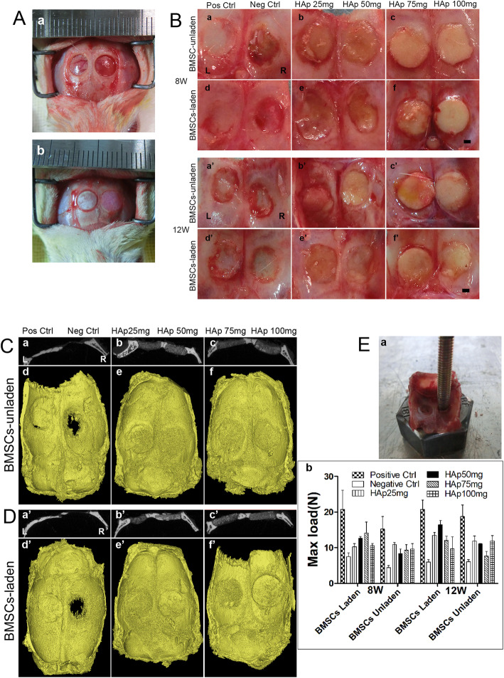
Fig. 5.
Establishment of the rat calvarial defect model and implantation of the different scaffolds into the defect. A (a) The calvarial defect model, where the diameter of the calvarial defect is 5 mm. (b) Autograft bone and PEG/SF/HAp scaffold were implanted into the calvarial defect. B The morphology of the regenerated tissues in the calvarial defect area after 8 and 12 weeks (inside view, scale bar = 1 mm). (a–c and a’–c’) BMSC-unladen positive control group, negative control group, HAp 25 mg group, HAp 50 mg group, HAp 75 mg group, and HAp 100 mg group, respectively. (d–f and d’–f’) BMSC-laden positive control group, negative control group, HAp 25 mg, HAp 50 mg, HAp 75 mg, and HAp 100 mg groups. C (a–c) Gray value of CT image of coronal plane calvarial defect repaired specimen. (d–f) 3D reconstruction images of micro-CT scanning data of the samples after 12 weeks of calvarial defect model by PEG/SF/HAp scaffold implanted with unladen BMSCs (inside view). D (a’–c’) Gray value of CT image of coronal plane calvarial defect repaired specimen. (d’–f’) 3D reconstruction images of Micro-CT scanning data of the samples after 12 weeks of calvarial defect model by PEG/SF/HAp scaffold implanted with laden BMSCs (inside view). E Biomechanical properties of regenerated tissue. (a) Biomechanical testing process. (b) The maximum load force applied to the calvarial defect area repaired by scaffolds

