Figure 5.
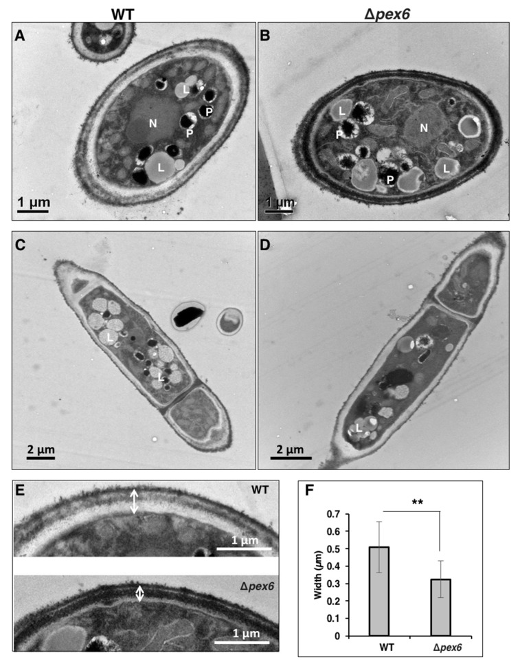
Transmission electron microscopy of A. alternata hyphae. (A) Cross section of a hypha of wild type (WT); (B) Cross section of a hypha of a pex6 mutant (Δpex6); (C) Longitudinal section of a hypha of WT; (D) Longitudinal section of a hypha of Δpex6. Abbreviations: N, nucleus; P, peroxisome; L, lipid body; (E) Images of fungal cell walls; (F) Measurement of cell-wall thickness. Means (n = 30) indicated by asterisks are significantly different from one another, p < 0.01 (**).

