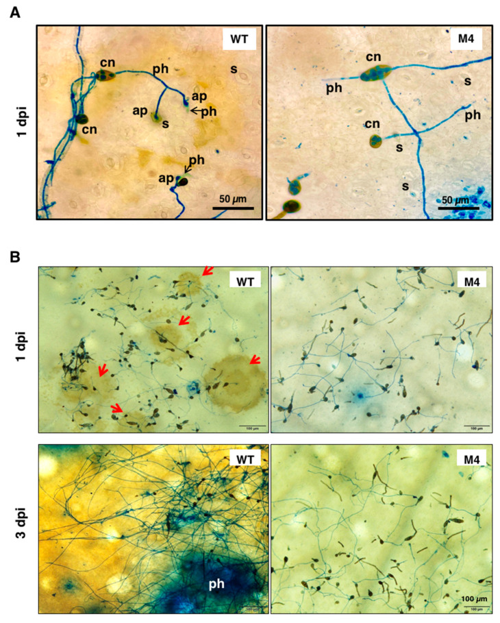
Figure 7.
Light microscopy observation of conidia germination on the surface of calamondin leaves. (A) Conidia of wild type (WT) and the Δpex6-M4 mutant were placed on citrus leaves and observed by light microscopy, revealing the germination of conidia (cn), the formation of appressorium-like structures (ap) and the penetration of hyphae (ph) through stomata (s) after being staining with lactophenol cotton blue; (B) Microscopic lesions (indicated by red arrows) were observed on the surface of citrus leaves inoculated with wild-type conidia 1 day post inoculation (dpi) and coalesced to become large necrotic lesions 3 dpi. Microscopic lesions were rarely observed on citrus leaves inoculated with conidia collected from the Δpex6-M4 mutant.

