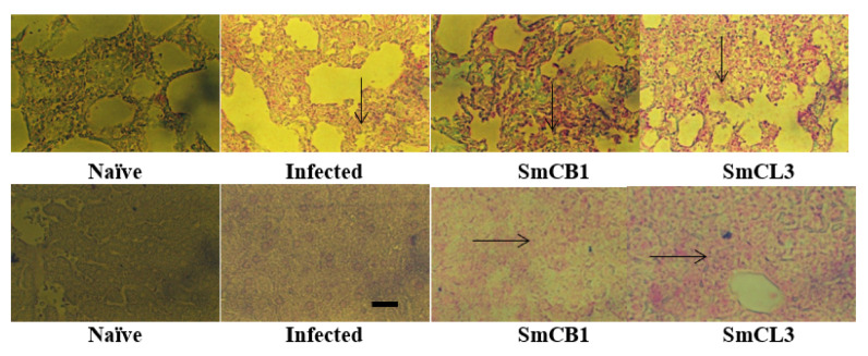
Figure 4.
Lung (upper panel) and liver (lower panel) of each of two naïve, unimmunized (Infected) and SmCB1-(SmCB1) and SmCL3-(SmCL3) immunized mice were reacted with irrelevant control (Supplementary Material, Figure S3) or anti-ARA antibody (Ab, MBS2003715, MyBioSource, San Diego, CA, USA), then alkaline phosphatase-labeled antibody to rabbit immunoglobulins and the reaction was visualized with Histomark RED Phosphatase Substrate Kit of Kirkegaard and Perry Laboratories. The arrows point to the areas of intense reactivity. The images shown are representative of the consistently recorded reactivity for each mouse group on day 10 pi (upper panel), and day 17 and, 24 (lower panel post S. mansoni infection. ×200; scale bar = 20 μM.

