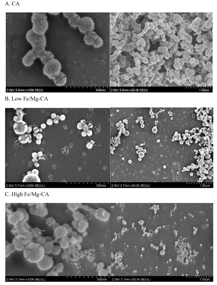
Figure 7.
FESEM micrographs in presence of 10% FBS showing size, shape and surface morphology. Scale bar: 500 nm, and 1 µm. (A) CA (B) Low Fe/Mg-CA (C) High Fe/Mg-CA. After the respective particles were incubated and supplemented with 10% FBS, they were centrifuged twice for 15 min at 13,000 rpm. The supernatants were discarded and the pellets were resuspended in 20 µL Milli Q water. Then 3 µL of the respective NP suspensions was dried at room temperature for 45 min.

