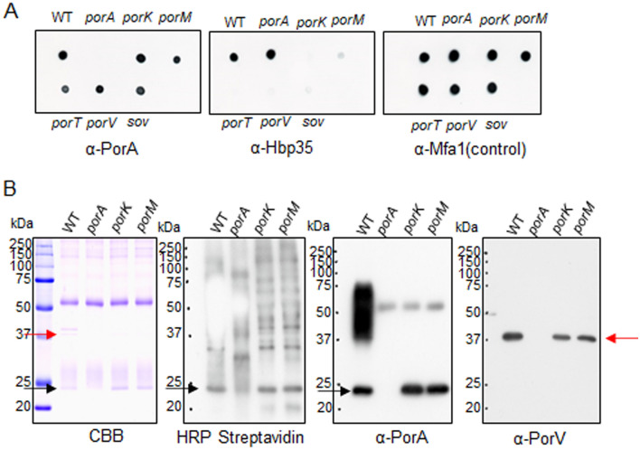
Figure 3.
Cell surface localization of PorA in P. gingivalis cells. (A) Cells of P. gingivalis wild type, ΔporA, porK, porM, porT, porV, and sov strains were blotted on nylon membranes and immunodetected by antibodies against PorA (α-PorA), Hbp35 (α-Hbp35), and Mfa1 (α-Mfa1). (B) Biotin-labeled cells of P. gingivalis wild type, ΔporA, porK, and porM strains were lysed and then immunoprecipitated by α-PorA. The immunoprecipitated samples were subjected to SDS-PAGE, followed by immunoblot analyses using HRP conjugated streptavidin, α-PorA, and antibody against PorV (α-PorV). CBB: Coomassie Brilliant Blue staining. Red and black arrows indicate PorV and PorA proteins, respectively.

